Have you ever wondered how dentists perform treatments and surgeries precisely and accurately inside our mouths?
Dentists use unique dental equipment called oral X-rays, which enables them to determine the condition of your teeth and curate specific treatment plans.
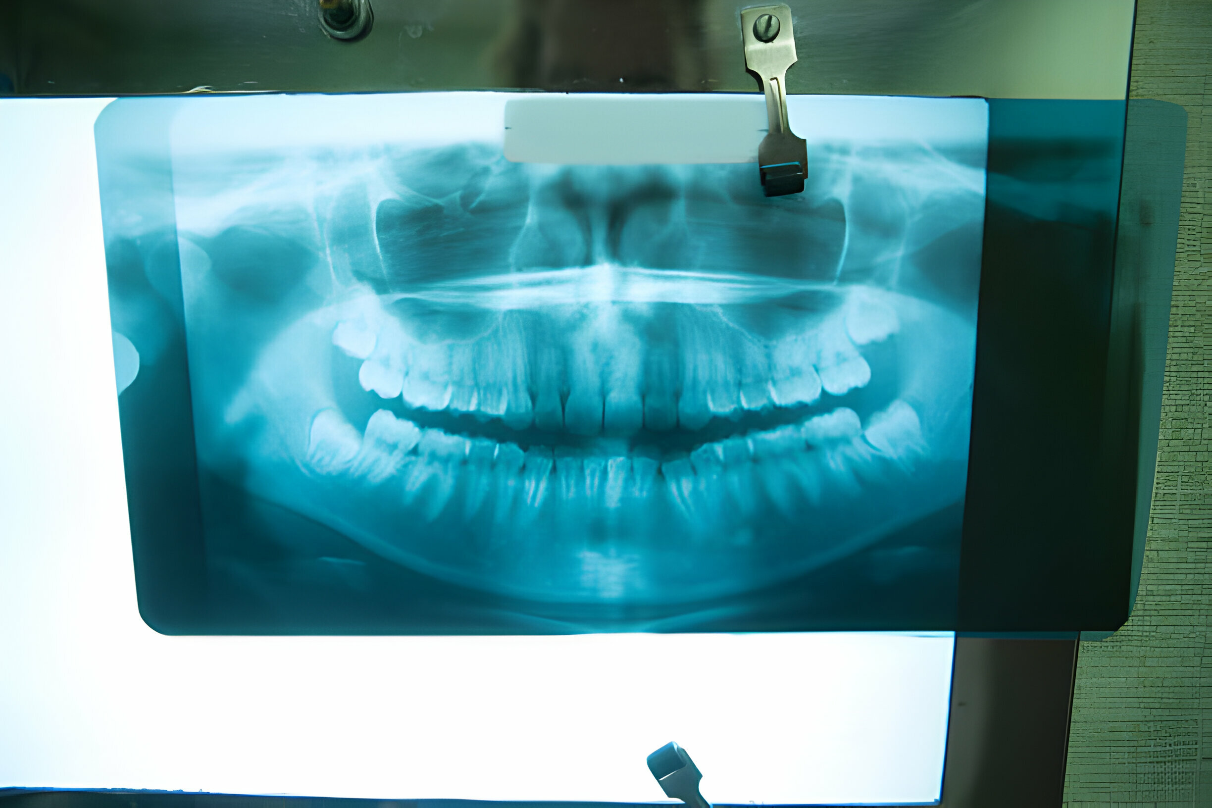
Dental issues are common among every age group; sometimes, most problems aren’t visible or noticeable. That’s when they use X-rays from your professional Dental Summerville, SC, to determine the condition of your oral health for further treatment procedures. But how many X-rays are there, and what are they used for?
In this blog, we shall delve into topics like:
Continue reading as we explore different types of dental X-rays and their uses in the following sections.
Dental X-rays, or radiographs, are invaluable tools in dentistry, allowing dentists to see beyond what is visible to the naked eye during a regular examination. They play a crucial role in diagnosing various conditions such as cavities, gum disease, and impacted teeth by providing detailed images of the teeth, roots, and surrounding bone structure.
Dentists rely on Summerville Dental X-rays to create precise treatment plans tailored to each patient’s needs, ensuring effective management of problems despite concerns about radiation exposure. Modern X-ray technology minimizes risks while maximizing diagnostic accuracy, ultimately contributing to better oral health outcomes for patients.
Dental X-rays, the unsung heroes of oral healthcare, serve as indispensable tools in the arsenal of modern dentistry. They unveil hidden aspects of oral health, revealing conditions that might elude the naked eye during routine examinations.
Let’s understand the different types of oral X-rays, each type a distinct window into the intricate world of oral anatomy and pathology.
Imagine a snapshot capturing the intricate dance of molars and premolars, revealing the subtle nuances of interproximal spaces. Bitewing X-rays excel in detecting the earliest signs of decay between teeth, an area notoriously challenging to assess visually. They also offer insights into the integrity of restorations, ensuring that fillings and crowns snugly embrace their designated territories.
Delving deeper into the cavity, periapical X-rays zoom in on individual teeth, from crown to root apex. Like archaeological excavators, they unearth hidden mysteries lurking beneath the gumline.
These X-rays play a pivotal role in diagnosing conditions such as abscesses, root fractures, and anomalies in tooth morphology. Periapical X-rays guide clinicians in formulating targeted treatment strategies by providing a holistic view of the tooth’s structure and surrounding bone.
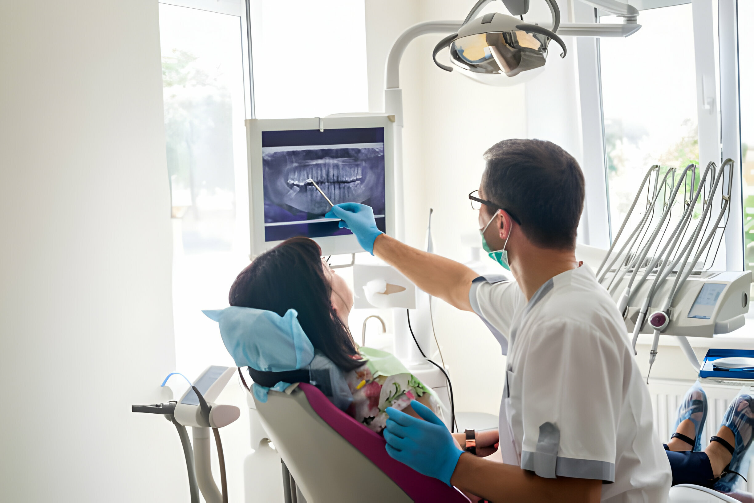
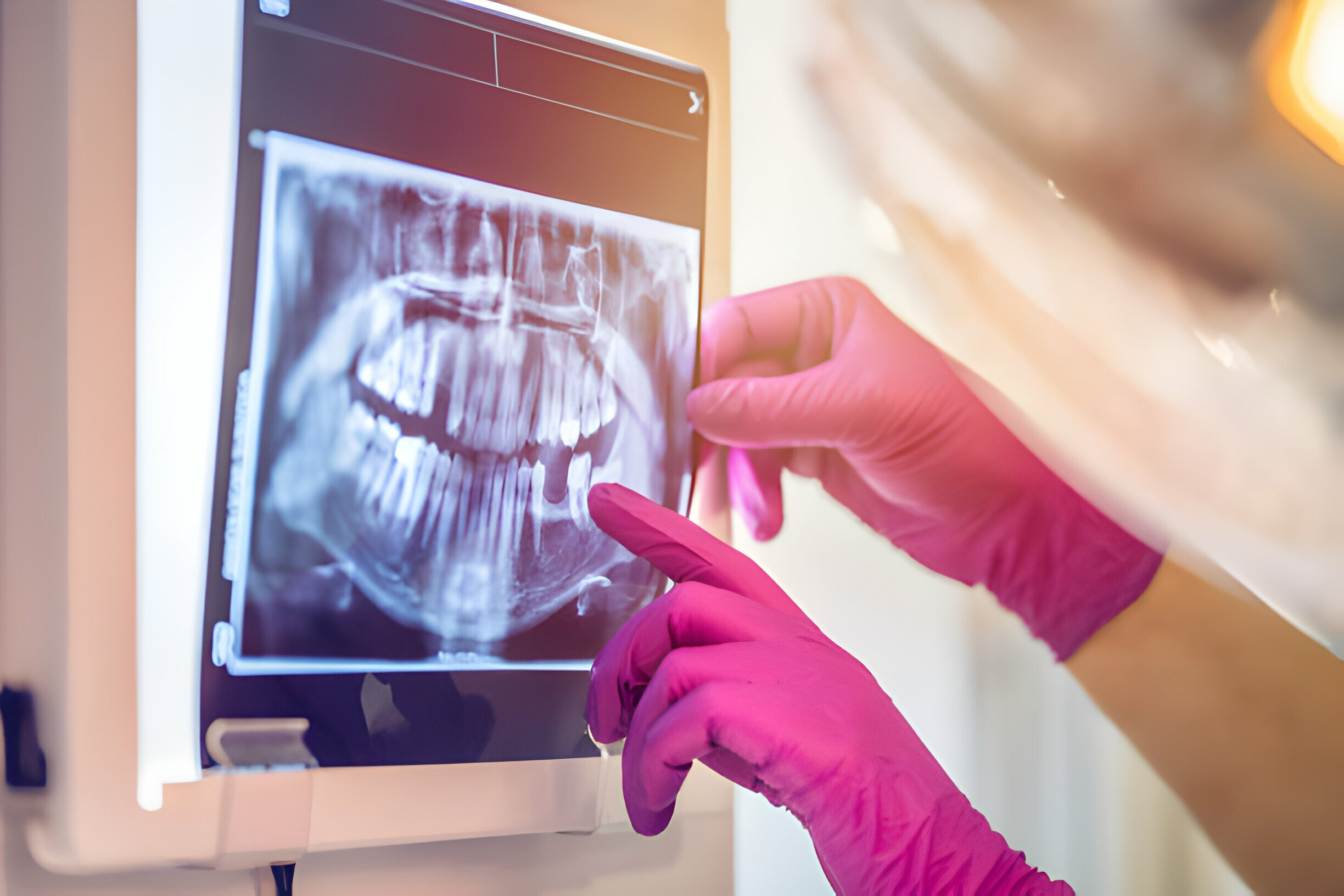
If X-rays were canvases, panoramic X-rays would be masterpieces, capturing the panoramic vista of the oral cavity in a single frame. They encompass the teeth, jaws, sinuses, and temporomandibular joints with a sweeping gaze.
Panoramic X-rays are indispensable allies in orthodontic planning, detecting impacted teeth, and evaluating the development progression. They offer a panoramic perspective, illuminating the broader landscape of oral health.
In orthodontics, precision is paramount, and orthodontic X-rays rise to the occasion with finesse. Also known as cephalometric X-rays, they shift the focus to the side profile of the head, unraveling the intricate interplay between teeth, jaws, and facial structures.
Orthodontic X-rays serve as blueprints for treatment, aiding orthodontists in assessing dental alignment, diagnosing skeletal discrepancies, and crafting personalized treatment plans tailored to each patient’s unique anatomical landscape.

Enter the realm of three-dimensional imaging, where anatomy springs to life with unparalleled clarity and precision. CBCT, the crown jewel of radiology, revolutionizes diagnostic capabilities with its ability to generate high-resolution 3D images of teeth, jaws, and surrounding tissues.
From intricate dental implant placements to complex orthognathic surgeries, CBCT guides clinicians with surgical precision, minimizing guesswork and maximizing treatment outcomes.
As the curtain falls on our exploration, occlusal X-rays take center stage, illuminating the hidden recesses of the oral cavity’s floor. With a broad sweep, they unveil the entire arch of teeth, unveiling anomalies such as cysts, tumors, or supernumerary teeth.
Occlusal X-rays serve as invaluable tools in pediatric dentistry, offering insights into the development of primary teeth and detecting abnormalities that might impact oral health and development.
Dental x-rays are safe when you follow proper precautions. These machines require minimal radiation exposure. It exposes a person to at least 2-3 REM compared to the 360 REM that people usually get exposed to from the environment yearly. The benefits of timely detection and treatment of oral problems generally outweigh the associated risks of x-ray imaging.
Dental X-rays serve as indispensable tools in the realm of oral healthcare, offering many benefits that contribute to the well-being of patients. Here’s a closer look at why X-rays are an essential component of dental practice: